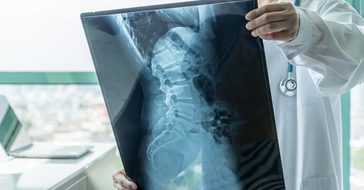
Radiography is the X-ray based method of photographing the inside of the body. These images let doctors view organs, bones, and other areas without surgery. It enables fast and safe discovery of injuries or infections.
Has anyone shattered a bone and undergone an X-ray? That radiography is in motion. It provides doctors with the answers they require, is quick and painless. These days, it finds employment in dentist offices, hospitals, and even airports.
This page will teach you the history of radiography, how it evolved over time, and the reasons it is so vital in the modern society. We will also discuss the instruments it employs and the several daily ways it benefits individuals.
The Science Behind Radiography
Radiography is a medical imaging method whereby images of the inside of the body are produced by use of radiation that of X-rays. Different tissues absorb X-rays differently as they transit through the body. Dense materials like bones absorb more X-rays and show up on the image white. Like organs and muscles, softer tissues absorb less X-rays and seem to be in greyscale. Doctors can see and diagnose diseases such as fractures or infections by means of this contrast.
An X-ray machine focusses a regulated beam of radiation at the area of interest in radiography. A detector gathers the X-rays passing through the body and turns them into a picture. After then, one can examine this picture on a computer screen. The short, non invasive procedure gives doctors useful information to help them to make correct diagnosis and create suitable treatment plans.
You May Also Read This Blog: Durable and Safe Hospital Stretchers for Patients
How Radiography Works: Principles of X-Ray Imaging
Radiography uses an X-ray beam sent through the body. Together, the X-ray tube, generator, and detector grab an image on a screen. This picture features varyingly coloured organs, tissues, or bones. In clinical diagnostics and diagnostic medicine, it aids in clinicians’ speedy and unambiguous decision making.
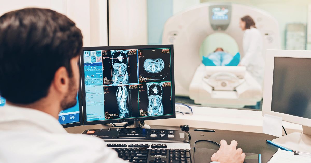
In hospital imaging departments and outpatient imaging centres, X-ray imaging is among the most reliable imaging techniques applied. Image quality rises and radiation exposure stays low with digital imaging. Many instruments also assist: Computed Tomography (CT), ultrasonic, magnetic resonance imaging (MRI). From radiologic equipment service to preventative equipment maintenance to AI driven diagnostics, these tools offer safer treatment and improved patient outcomes all throughout Pakistan.
Types of Radiography
Many imaging modalities found in radiography support diagnosis in medicine. Most often employed in hospital imaging departments and outpatient imaging facilities is X-ray imaging. It catches clear body images using an X-ray tube, detector, and generator. With minimum radiation exposure, this technique facilitates digital imaging and enhances X-ray image quality.
Advanced kinds of radiographic imaging are computed tomography (CT) and contrast imaging. In clinical diagnosis, CT scans provide cross sectional imaging to assist physicians. Dyes help contrast imaging reveal organs and blood flow. Supported by healthcare technology, AI driven diagnoses, and preventative equipment maintenance, these instruments enhance patient care and let service providers provide Pakistan with better healthcare imaging solutions.
Role of Radiation in Producing Diagnostic Images
Radiography produces clear images of the inside of the body by means of a low dosage of radiation. The X-ray tube sends photons through the body, where a detector catches them. Key components of diagnostic medicine and clinical diagnostics, X-ray imaging is made possible in part by this mechanism.
Computed tomography (CT) scans and modern digital radiography systems both leverage healthcare technology to raise picture quality while maintaining radiation safety under control. Radiographic imaging has gotten faster and safer with features including smart software systems, remote monitoring, and AI driven diagnostics. These medical imaging technologies help Pakistani hospital imaging departments, outpatient imaging centres, and improve patient care overall.
Safety Measures and Radiation Risks in Radiography
Although radiography is used in diagnosis, it also entails radiation exposure. Radiation safety is thus really crucial. To lower dangers, trained imaging experts set the X-ray tube, generator, and detector in safe environments. Following regulatory compliance helps digital radiography equipment safeguard staff members and patients in hospital imaging departments.
Service experts in Pakistan avoid overexposure by means of preventative equipment maintenance and remote monitoring. Other organisations such as Radiological Service Training Institute (RSTI) provide medical equipment certification and imaging service training. These initiatives preserve patient care, enhance image quality, and reduce clinical diagnostics risk. Every day, radiographic imaging is also becoming even more safer thanks to new healthcare technologies and AI driven diagnoses.
Key Applications of Radiography in Healthcare
By enabling clinicians to view within the body without surgery, radiography is becoming important in diagnosis medicine. It aids clinical diagnostics at imaging centres, outpatient and hospital imaging departments. Computed Tomography (CT) and digital X-ray devices assist identify early on cancer, lung infections, and bone fractures.
More choices for accurate and safe treatment come from other imaging modalities including ultrasound and magnetic resonance imaging (MRI). These devices reduce radiation exposure while yet improving patient outcomes and image quality. Supported by organisations like Radiological Service Training Institute (RSTI), trained imaging specialists and service professionals in Pakistan keep high equipment reliability by means of preventative equipment maintenance and imaging system maintenance.
Diagnostic Imaging
Radiography and diagnostic imaging enable clinicians to early on identify tumours, infections, and fractures. For quick and clear answers it employs X-ray imaging, computed tomography (CT), and ultrasound. Working in hospital imaging departments and outpatient imaging facilities, these imaging modalities serve to improve patient outcomes all over Pakistan’s healthcare system.
Doctors examine organs, bones, and tissues clearly thanks to digital radiography devices and clinical diagnostics instruments. Image quality is enhanced and service downtime is lowered by technologies including AI driven diagnostics and remote monitoring as well as by the support of qualified imaging experts. Radiation safety guidelines guide imaging diagnostics to safeguard individuals undergoing testing.
Preventive Screening
Preventive screening depends much on radiography since it enables doctors to early stage illness detection. Examining the body it employs X-ray imaging, ultrasonic, magnetic resonance imaging (MRI), and computed tomography (CT). By means of early therapy, these imaging technologies promote diagnostic medicine and enhance patient outcomes.
Faster results and improved image quality are achieved by Pakistani hospitals using digital radiography systems nowadays. Services follow radiation safety guidelines under the Radiological Service Training Institute (RSTI) with the help of qualified imaging experts. Advanced healthcare technologies and regular routine maintenance help to lower radiation exposure and enable robust healthcare imaging solutions.
Interventional Radiography
In interventional radiology, radiography is indispensable and enables doctors to treat patients without open operation. Doctors can securely direct equipment throughout the body using X-ray imaging, ultrasonic, computed tomography (CT), magnetic resonance imaging (MRI). These imaging techniques lower pain and recovery time, thereby enhancing patient care.
To do such operations, Pakistani hospitals employ qualified imaging experts and digital radiography technology. Strict radiation safety criteria and the Radiological Service Training Institute (RSTI) help clinicians guarantee improved clinical diagnosis. Using healthcare technologies, remote monitoring, and AI driven diagnostics helps to raise general patient outcomes and image quality.
Therapeutic Radiography
Fighting cancer requires radiography in major part. High energy radiation is applied in therapeutic radiography to destroy cancer cells and prevent their proliferation. By means of appropriate radiation safety, advanced X-ray imaging, computed tomography (CT), and magnetic resonance imaging (MRI) assist clinicians in carefully planning therapy and safeguarding of healthy tissues.
To treat cancer precisely, Pakistan’s hospitals apply digital radiography technologies, artificial intelligence driven diagnostics, and remote monitoring. Supported by Radiological Service Training Institute (RSTI) and knowledgeable imaging experts, patient care and image quality enhance. Every year, technical developments and healthcare technologies help to lower radiation exposure and boost beneficial patient results.
Common Tests
Radiography allows clinicians to quickly detect a range of medical problems. Chest X-ray is by far the most regularly used diagnostic tool in diagnosis oriented medicine. With X-ray imaging, one can see healthy or abnormal condition of the lungs, heart, and bones. Digital X-ray machines reduce patient waiting times while delivering higher quality images with reduced radiation.
The majority of major hospitals in Pakistan use abdominopelvic CT scans to find out if a patient has tumours, stones, or infections. In these procedures, CT scans and ultrasound are utilised for medical diagnosis. Because of the work of Radiological Service Training Institute (RSTI) and similar groups, patients receive superior outcomes thanks to imaging specialists and the use of AI for diagnosis.
Radiography Across the Body
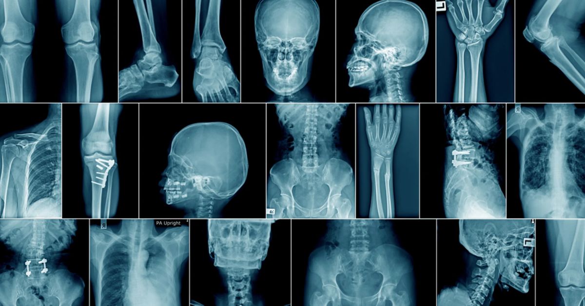
Radiography is the major approach used to assess the human body. Most often, X-ray machinery is relied on to look into the state of teeth, joints, and bones. Physicians can use this system to spot fractures, infections, and arthritis. Because digital radiography equipment gives off less radiation and produces sharper images, the benefits for patients are improved.
Assessment of soft tissues requires the use of computed tomography and magnetic resonance imaging as leading tools. Clinicians use these images in cross section to discover tumours, blood clots, and damage affecting organs. Integrating artificial intelligence with skilled imaging diagnosis lets doctors improve illness diagnosis and support improved patient care at medical imaging sites and hospitals.
Head and Neck
Head and neck area disorders are diagnosed in great part by radiography. Brain scans routinely employ magnetic resonance imaging (MRI) to find problems including tumours or injuries. Essential for diagnostic medicine, it provides high resolution pictures of soft tissues. Correct outcomes depend on it for imaging experts.
Computed tomography (CT) offers cross sectional pictures in head CT and neck CT scans that aid in the identification of blockages, infections, or injuries. Rapid diagnosis in an emergency calls for these imaging modalities. Radiographic imaging has advanced with medical imaging technology, therefore lowering radiation dose and guaranteeing better patient outcomes.
Chest
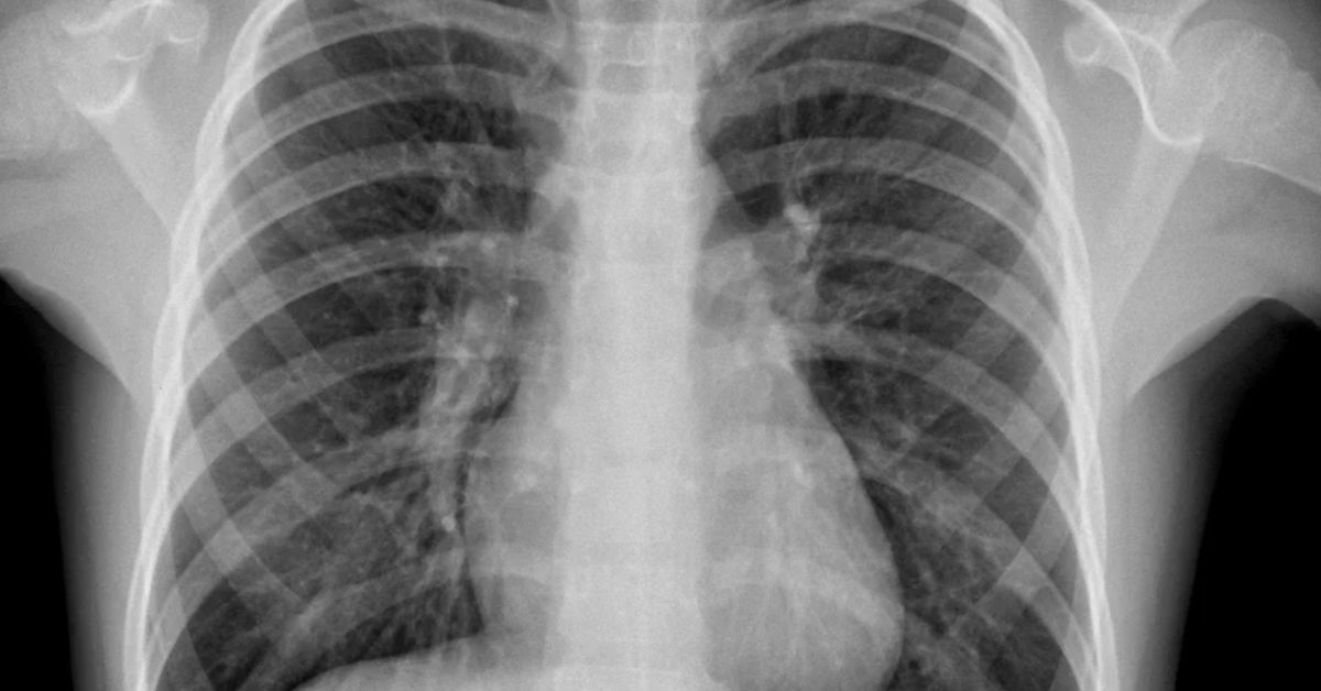
Diagnosing problems associated to the chest depends much on chest radiography. Commonly used imaging method for lung issues including infections or tumours is a chest X-ray (CXR). It is commonly available at imaging centres and outpatient clinics all throughout Pakistan, quick, non invasive. X-ray imaging lets doctors make rapid decisions.
Sometimes a more thorough inspection calls for a Chest CT scan. Clearer view of the lungs and chest organs is given by computed tomography (CT), which produces cross sectional images. Radiation safety is underlined with developments in medical imaging, so guaranteeing better patient outcomes and lower possible dangers.
Abdominopelvic Region
Diagnosing problems in the abdominopelvic area depends critically on radiography. Detailed cross sectional images from abdominopelvic CT scans enable doctors to find issues in organs including the liver, kidneys, and intestines. Commonly utilised in imaging centres and outpatient clinics all throughout Pakistan, this imaging technique is a basic instrument in diagnosis medicine.
Computed Tomography (CT) allows abdominopelvic CT to provide a better image of the inside body components. Conditions including cancer, infections, and injuries are found with this advanced imaging technology. Radiographic imaging guarantees excellent images with appropriate radiation safety precautions, therefore improving patient outcomes and enabling better diagnosis.
Upper Extremity
Determining problems with upper extremities depends critically on radiography. By giving doctors comprehensive pictures of the shoulder joint, shoulder MRI helps them to spot conditions such as bone fractures or rotator cuff tears. Through better treatment planning, magnetic resonance imaging (MRI) increases clarity, hence improving diagnosis, medicine, and patient care.
Wrist MRI provides good images for diagnosis of ligament injury, arthritis, and fractures in wrist injuries. MRI and other imaging technologies enable doctors to better estimate damage. This approach of medical imaging guarantees correct diagnosis, thereby improving patient outcomes and helping imaging experts to make wise treatment decisions.
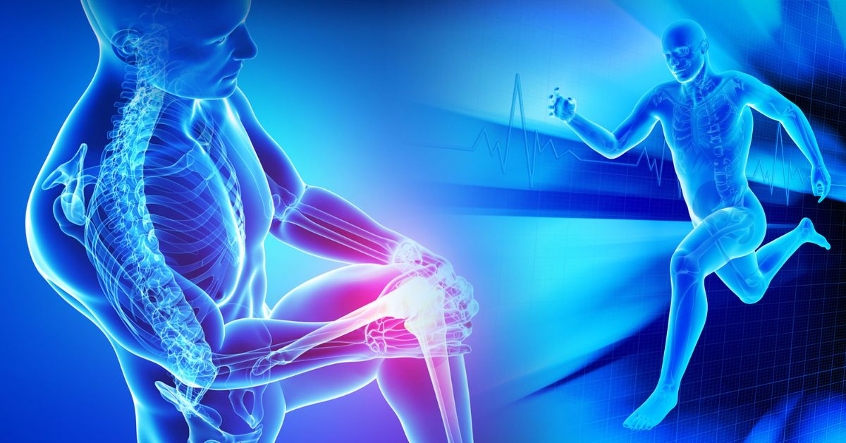
Lower Extremity
A large portion of diagnosing issues with the knee and ankle depends on using radiography techniques. Doctors mostly use magnetic resonance imaging (MRI) and X-ray imaging when diagnosing lower extremity issues. The use of these techniques generates clear images that identify fractures, ligament problems, and many additional issues, thereby improving both diagnosis and eventual patient outcomes.
Because MRI results show soft tissues more clearly, doctors rely on it to diagnose knee and ankle concerns. Owing to its detailed images, this process often spots injuries in the tendons or cartilage. Precise diagnosis and treatment decisions are supported by the use of MRI and CT scans in the imaging classification.
The Role of Radiographers and Radiologists
Medical experts called radiologists make images of the body using X-ray, MRI, and CT scanners among other imaging methods. Their efforts assist physicians in spotting and diagnosing illnesses or injuries. To safeguard patients and oneself, radiographers have to strictly abide by safety guidelines including radiation safety.
Radiologists are physicians that focus in reading the images produced by radiographers. To aid in diagnosis and treatment of medical disorders, they read X-ray, MRI, and CT scan findings. Their knowledge in imaging diagnostics guarantees correct diagnosis and directly influences patient treatment.
What Does a Radiographer Do? Responsibilities and Skills
Operating imaging equipment and ensuring that images are clear and correct fall to radiographers. They have to additionally guarantee patients’ comfort and get them ready for the operation. Technical knowledge of imaging technologies, patient care, and radiation safety is essential ability for radiographers. They are absolutely important in diagnosis medicine.
Radiographers must be excellent communicators to reassure patients and explain treatments. They also have to manage preventative equipment maintenance to guarantee it runs as it should. Programs designed to keep radiographers current on new radiographic equipment and imaging technologies are provided by the Radiological Service Training Institute (RSTI).
What Does a Radiologist Do? Interpreting Imaging Results
Radiologists specialise in reading radiographer produced medical images. To identify disorders including fractured bones or tumours, they examine X-ray, CT, and MRI scans. Determining the appropriate course of treatment for patients depends on their capacity to precisely understand visuals, therefore directly affecting patient outcomes.
To develop treatment regimens, radiologists often closely coordinate with other medical experts including oncologists and surgeons. Their decisions are supported by imaging modalities and cutting edge artificial intelligence driven diagnostics. High quality patient treatment depends on their knowledge in radiology and medical imaging.
Importance of Specialized Training for Radiography Professionals
Radiologists and radiographers alike depend on specialised training to guarantee they can run sophisticated imaging devices safely and successfully. From radiation safety to advanced digital imaging techniques, the Radiological Service Training Institute (RSTI) provides courses covering all aspects. Excellent imaging and low patient radiation exposure are guaranteed by appropriate training.
As healthcare technology develops, one must always be learning. Digital radiography equipment and other new imaging technologies call for service providers to be current. By generating better and more precise images, specialised training helps professionals adapt to these technological changes, therefore boosting their skills and patient care.
Adapting to Technological Advances in Radiography
Medical imaging technology’s developments including artificial intelligence driven diagnostics are revolutionising radiography. Radiologists and radiographers have to adjust to these developments to deliver the best treatment. Accurate diagnosis and effective imaging equipment use in hospital imaging departments depend on a knowledge of modern tools and software systems.
Remote monitoring and imaging system maintenance are among the technological developments that assist radiography equipment’s dependability to increase. Preventive maintenance guarantees that machines remain in good condition as digital X-ray systems proliferate, reducing downtime and ensuring excellent X-ray image quality for every patient.
Types of Radiology: A Broader Perspective

Radiography covers X-ray imaging, CT scans, and MRI among other imaging modalities. These imaging techniques enable physicians to examine within the body in search of diseases. Ultrasound is one of the instruments used in diagnostic medicine that lets doctors rapidly identify the reason of symptoms.
Every kind of radiation has special advantages for patient treatment. Bones are typically seen with X-ray, CT offers cross sectional views of organs. For soft tissues, MRI provides excellent pictures free of radiation. These methods together enable doctors to make accurate diagnosis, hence improving patient outcomes.
Diagnostic Radiology
Because it lets clinicians see inside body structures, diagnostic radiography is absolutely vital in healthcare. MRI, CT scans, and X-ray imaging among other methods offer finely detailed pictures of organs, tissues, and bones. These imaging techniques enable the identification of diseases, therefore supporting accurate diagnosis and efficient patient treatment.
Radiographic imaging is a tool used in diagnostic medicine to assist in the underlying cause of symptoms’ identification. Digital radiography presents reduced radiation exposure and better images. Early disease detection supported by imaging diagnostics enhances patient outcomes by means of early intervention. Furthermore, radiological service training guarantees imaging experts’ safe and effective use of modern technologies.
Interventional Radiology
Medical imaging is used in interventional radiology to direct surgeons during procedures. Often used for these treatments are X-ray imaging, CT, and MRI. This lets doctors treat disorders with few incisions, hence optimising healing periods and lowering risk of consequences. Safety and reliable results depend much on imaging experts.
Interventional radiology targets particular bodily locations by means of imaging modalities. Ultrasounds, for instance, can guide a needle removing fluid or doing a biopsy. Modern diagnosis depends on these image guided procedures, which enhance patient care and outcomes while lowering radiation exposure and recuperation times.
Therapeutic Radiology
Cancer treatment, in particular, depends on radiation based therapies in therapeutic radiology. X-ray and radiographic imaging methods help health professionals focus treatment accurately on cancerous tissues. Using advanced equipment including MRI and CT, clinicians can easily define the locations that need therapy, ensuring both maximum accuracy and protection against damage to normal tissue.
Radiation safety is of the greatest importance in therapeutic radiology to protect doctors and patients from harm. Careful attention must be paid to treatment techniques, even when they involve advanced imaging technologies. Image specialists are responsible for applying the right safeguards, which guarantees both good treatment and protection against unnecessary radiation.
Nuclear Medicine Imaging and Ultrasonography
Internal organs are seen in nuclear medicine imaging using radioactive materials. Especially for the diagnosis of cancer and heart disease, this approach offers crucial information. Conversely, ultrasonic waves record real time images using sound waves, which makes them useful for soft tissue evaluation for disorders including pregnancy and stomach problems.
Together, nuclear medicine and ultrasonic imaging provide a whole view of a patient’s health. Complementing these modalities, diagnostic instruments including X-ray imaging and CT scans offer more finely detailed findings. When radiation exposure is controlled, these imaging methods are safe and guarantee great image quality and enhanced patient outcomes.
Why Radiography is Vital to Healthcare

By offering accurate and crisp images of inside body structures, radiography is vital in diagnosis medicine. Early diagnosis of diseases is made possible by these medical imaging technologies, which include X-ray imaging and computed tomography (CT), therefore facilitating successful therapy. Early diagnosis helps patients far better.
Apart from diagnosis, radiographic imaging informs non surgical as well as surgical treatments. X-ray systems track illness development, technologies include magnetic resonance imaging (MRI) and ultrasonic help in therapy planning. Constant technical development enhances image quality, training under organisations like the Radiological Service Training Institute (RSTI) guarantees the safety and efficacy of these imaging systems.
Advances in Radiography Technology
Over the years, radiography technology has developed remarkably. From analogue to digital X-ray technology, it has evolved to better image quality and faster and more accurate diagnosis making. Accurate diagnosis imaging and treatment planning in healthcare technology depend on clear images, which digital X-ray systems provide.
Radiography has lately evolved with the application of machine learning and AI driven diagnosis. These developments contribute to raise image quality and radiation safety. Imaging modalities like CT and MRI as well as systems like PACS (Picture Archiving and Communication System) help clinicians make more exact decisions in patient care and treatment. Technological developments keep changing the discipline of radiologic education and clinical diagnostics.
Preparing for a Radiography Procedure
Patients should follow some basic guidelines to guarantee correct findings before to having a radiography treatment. Should you be undergoing an X-ray or CT scan, you may have to remove metal based clothes or jewelry. To guarantee your safety throughout the operation, tell your doctor of any past operations or medical issues.
The radiologic technologist will properly position you for clear radiography during the treatment. To produce finely detailed images, imaging facilities employ CT scanners and sophisticated digital X-ray systems. Staying steady during the scan will help you to avoid fuzzy pictures. Most procedures cause little discomfort and are short and non invasive.
What to Expect During a Radiography Procedure
Usually starting a radiography process with a review of your medical history helps to evaluate any dangers. You might be requested to lie on a table for a CT scan or X-ray imaging as the X-ray tube, detector, and generator cooperate to produce clear images. The technologist will walk you through every phase.
Some operations call for the use of contrast materials to improve pictures, which can entail injection or swallowing. Safety from radiation comes first always, particularly in delicate situations like pregnancy. A radiologist will examine the pictures after the operation and forward the findings to your doctor.
Understanding Contrast Materials: Uses and Precautions
In radiography, contrast materials are agents that make particular body parts more conspicuous during imaging. Such materials are used during CT scans and X-rays to better show the shape of internal organs or blood arteries. Contrast materials are occasionally given by injection, sometimes orally, or occasionally by alternative ways into the body, all depending on what the operation demands.
Though contrast materials are generally without risk, people with allergies or kidney problems should take extra precautions. You ought to inform your doctor of your medical history just before the operation. Minor adverse reactions from contrast materials include sensations of warmth or a brief metallic flavor. Your safety is best maintained by carefully following your healthcare practitioner’s instructions.
Radiography in Pregnancy: Safety Considerations
Radiography taken during pregnancy calls for special attention. Tell your radiologic technologist whether you are pregnant or might be since radiation exposure during pregnancy might cause hazards. With appropriate shielding and care, many imaging treatments are safe.
Sometimes non radiation imaging choices such as MRI or ultrasonic waves could be taken under consideration as substitutes to guarantee the mother’s and the baby’s protection. Open communication with your healthcare practitioner should always come first in order to make wise decisions on the best line of action during pregnancy.
Understanding Radiography Results
Detailed images of the interior of your body produced by radiography enable doctors to recognize abnormalities. Standard procedures make use of X-ray images, as well as CT scans and MRI. Such results may reveal organ abnormalities, infections, forms of cancer, or fractures. Following image assessment, the radiologist updates your doctor about what they found.
Your results will be available after imaging according to the demands of the particular type of scan used. In most cases, results from basic X-ray scans will be ready in just a few hours. Because specialized equipment and careful analysis are essential for CT and MRI examinations, the results from these operations may take longer than X-rays.
What to Expect During Radiography Results Review
Should your radiography findings be normal, you might not have to proceed any more. If aberrant results are discovered, nevertheless, your doctor will go over with you the following actions. To verify a diagnosis and choose the best course of action, this can call for more testing or imaging scans.
Should your doctor recommend additional diagnostic instruments or treatment, it is advisable to perform as advised. A follow up visit or specialist treatment could be required for aberrant findings. If you have questions or require explanation on the radiography findings, always see your doctor.
Risks and Side Effects of Radiography
X-ray imaging, CT scans, and other diagnostic techniques are used in radiography to provide finely detailed body images. Although the diagnosis of many health disorders advantages are great, it is crucial to be aware of the possible hazards. Particularly when utilized regularly, radiation exposure from these imaging techniques can cause long term health issues to be worried about. Still, the existing radiation safety precautions greatly lower these hazards.
Sometimes radiographic imaging uses contrast materials to improve the quality of the pictures. Although these drugs are usually safe, adverse effects including kidney issues or allergic responses might occur. Reducing any possible hazards mostly depends on following the radiation safety recommendations and lowering exposure. Frequent maintenance of imaging systems guarantees effective operation of equipment, therefore lowering risks.
Minimizing Risks Through Best Practices
Imaging experts use best practices, such employing the lowest feasible radiation exposure for the necessary image quality, to lower the hazards related with radiographic equipment. Digital radiography systems’ and other equipment’s preventive maintenance guarantees their safe operation. Furthermore, frequent service training courses support experts in diagnostic medicine to keep high safety standards.
Those who require regular imaging that is, those with chronic diseases should give great thought to the long term hazards. Safer imaging methods and AI driven diagnostics are driving ongoing evolution in healthcare technologies. Reducing pointless scans and concentrating on clinical diagnostics help to control possible health issues while nevertheless provide vital diagnostic data.
The Future of Radiography
New technology are fast changing radiography. More exact and detailed images made possible by 3D imaging are enhancing diagnosis possibilities. This development improves radiographic imaging for MRI and CT scans among other fields. Faster outcomes and more exact diagnostics are the direction medical imaging is headed.
AI driven diagnostics’ integration is transforming diagnosis based treatment. Better patient outcomes follow from AI helping to lower human error and improve accuracy. Programs for imaging experts’ service training are really vital if we are to support these advancements. These initiatives guarantee radiation safety and imaging quality while making radiographers ready to apply modern technologies.
FAQ’s
What is radiography of the spine?
Using X-rays, radiography of the spine produces images of the spine’s bones and structures to identify conditions such fractures or alignment abnormalities.
What is a radiography in medical imaging?
In medical imaging, radiography is the technique whereby X-rays are used to provide finely detailed images of the inside of the body for disease diagnosis.
What is radiographic imaging for skeletal system?
Radiographic imaging for the skeletal system is the visualizing of bones and disease, fracture, or anomaly detection using X-rays.
What is the function of the radiography?
Radiography serves to provide images of inside body structures, therefore enabling doctors in diagnosis and treatment of medical disorders.
What is the principle of radiography?
Radiography is the application of X-rays passing through the body to create images on a film or digital detector thereby visualizing internal structures.
Conclusion
By allowing doctors to identify and treat a wide spectrum of diseases, radiography is absolutely crucial in healthcare. It offers precise, detailed images to help with early identification and prevention. Radiography helps people to uncover possible problems before they become major by means of proactive health screening. Maintaining knowledge about the accessible radiography techniques helps one to guarantee best health and well being. Discover today how radiography might improve your health and help to support better medical results.